Sterile Laboratory Reconstitution, Solvent Selection & 0.22 μm Filtration
Dissolving a freeze-dried lyophilized peptide cake into an aqueous stock solution is a critical physicochemical transition. Selecting a solvent near the peptide's isoelectric point (pI), introducing high-velocity fluid shear, or filtering through high-binding nylon or cellulose nitrate membranes can precipitate up to 60% of the active peptide out of solution or induce irreversible beta-sheet fibril aggregation.
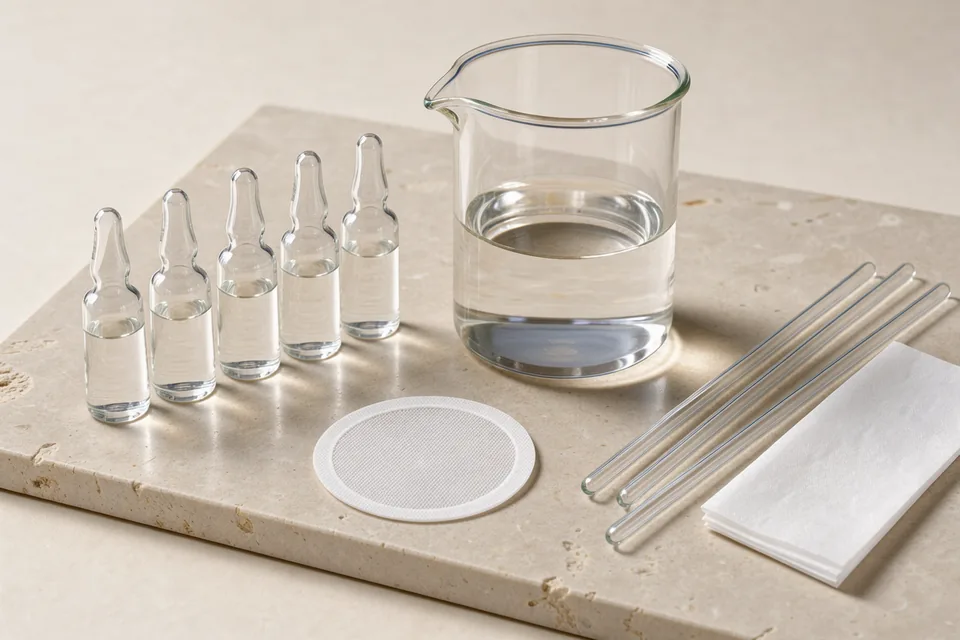
Matching Reconstitution Solvent to Net Formal Charge & Hydrophobicity
Before introducing any aqueous buffer to a lyophilized vial, calculate the net formal charge (Z) of the amino acid sequence at pH 7.0 by summing positively charged basic residues (Arg, Lys, His, and unblocked N-terminus) against negatively charged acidic residues (Asp, Glu, and unblocked C-terminus).
| Peptide Physicochemical Class | Primary Laboratory Solvent | Chemical Mechanism & Compatibility | Incompatibilities & Assay Caveats |
|---|---|---|---|
| Hydrophilic / Polar Sequences (≥25% charged residues; net charge Z ≠ 0) |
Sterile Type I Ultrapure Water (18.2 MΩ·cm) | Deionized, endotoxin-free (<0.005 EU/mL) water dissolves lyophilized TFA or acetate peptide salts rapidly without introducing competing salt ions that cause "salting-out" prior to complete hydration. | Unpreserved sterile water supports microbial growth if opened repeatedly outside an ISO Class 5 laminar flow biosafety cabinet; single-use cryo-aliquoting at -80°C is mandatory. |
| Multi-Aliquot Bench Reference Stocks (Non-cell-culture analytical standards) |
0.9% Benzyl Alcohol Bacteriostatic Water | Contains 0.9% w/v (9 mg/mL) benzyl alcohol as an aromatic bacteriostatic preservative, inhibiting microbial proliferation in multi-withdrawal analytical calibration standards stored at 2°C to 8°C. | HPLC & Cell Culture Caveat: Benzyl alcohol absorbs intensely at 214 nm and 254 nm (producing a massive solvent peak on RP-HPLC) and is cytotoxic to primary neuron, hepatocyte, and stem cell cultures. |
| Basic / Polycationic Peptides (Net charge Z > +1; rich in Lys, Arg, His; e.g., SS-31, LL-37) |
Dilute Acetic Acid (0.01% to 0.1% v/v Aqueous HOAc, pH 3.5–4.8) | Protonates basic side chains (especially Histidine, pKa ≈ 6.0), maximizing electrostatic repulsion between peptide chains and preventing isoelectric precipitation at neutral pH. | Avoid adding concentrated PBS directly to dry basic peptides containing residual TFA or acetate counterions until primary dissolution in dilute acid or water is complete. |
| Acidic / Polyanionic Peptides (Net charge Z < −1; rich in Asp, Glu; e.g., acylated GLP-1/GIP sequences) |
Dilute Ammonium Hydroxide or 10–50 mM Sodium Bicarbonate (pH 7.8–8.4) | Deprotonates carboxylic acid side chains (Asp, Glu) above their pKa (~4.0), generating soluble carboxylate anions (-COO−). | Do not acidify polyanionic sequences below pH 5.0; crossing the isoelectric point (Z = 0) triggers immediate turbid flocculation and irreversible beta-sheet gelation. |
Sterile 0.22 μm Membrane Filtration: PES & PVDF vs. Nylon
When preparing sterile peptide stock solutions for primary cell culture, organoid perfusion, or receptor pharmacology assays inside a biosafety cabinet, terminal filtration through a 0.22 μm sterile syringe filter removes vegetative bacteria and sub-visible particulates. However, filter membrane polymer chemistry dictates whether the peptide passes into the filtrate or binds irreversibly to the filter housing:
- Hydrophilic Polyethersulfone (PES) & Polyvinylidene Fluoride (PVDF) — Recommended: Low-protein-binding hydrophilic PES and PVDF membranes exhibit non-specific peptide adsorption below 1.5 μg/cm². For small-volume stocks (1.0 mL to 5.0 mL), select a 4 mm or 13 mm diameter syringe filter (hold-up volume <15 μL) rather than a 25 mm filter (which retains >100 μL of dead volume).
- Nylon (Polyamide) & Cellulose Nitrate — Strictly Avoid: Nylon and mixed cellulose ester (MCE) membranes carry abundant hydrogen-bonding amide and nitro groups that avidly bind amphipathic and hydrophobic peptides, stripping 30% to 75% of a dilute (<100 μg/mL) peptide stock out of solution during a single pass.
Preventing Peptide Aggregation, Foaming & Mechanical Shearing
Synthetic polypeptides—particularly amphipathic helices and beta-hairpin structures—behave as surface-active surfactants in aqueous solution. At the air-water interface of a microbubble, hydrophobic amino acid side chains orient toward the air phase while hydrophilic residues remain in the water phase, unfolding the native conformation and nucleating insoluble amyloid-like fibrils:
- Thermal Equilibration Prior to Septum Puncture: Remove the sealed lyophilized vial from -20°C storage and equilibrate in a desiccator at ambient room temperature (20°C–22°C) for 20 minutes before introducing solvent. Puncturing a cold vial causes immediate condensation of atmospheric moisture onto the hygroscopic cake.
- Laminar Wall-Wet Introduction: Direct the solvent stream gently down the inner borosilicate glass wall rather than jetting liquid directly into the lyophilized cake. High-velocity fluid impingement creates micro-aerosols and air-water interface foaming that denature amphipathic sequences.
- Zero Vortexing or Ultrasonic Agitation: Never place a reconstituted peptide vial on a high-speed mechanical vortex mixer or in an ultrasonic bath. Cavitation and high-shear turbulence induce hydrodynamic shearing and disulfide scrambling. Dissolve the cake exclusively via gentle orbital swirling for 60 to 120 seconds until optically clear.
Related Technical Manuals: Review our Lyophilized Solvent Dissolution & Buffer Chemistry Monograph, Thermal Degradation Kinetics & Cryogenic Storage, and USP <85> Bacterial Endotoxin Verification.